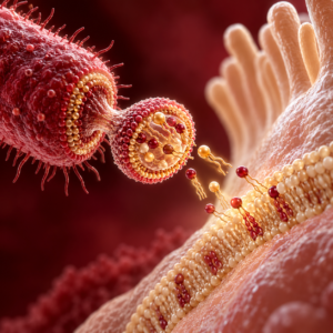
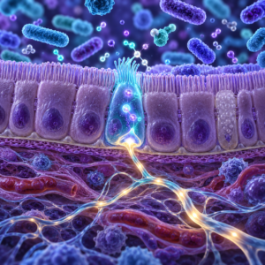
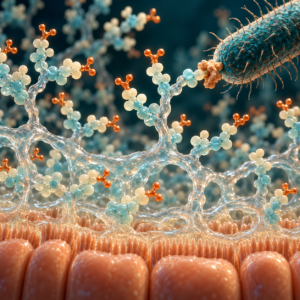
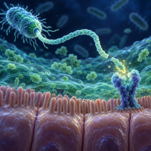
The vagus nerve is often described as a telephone line between gut microbes and the brain. That comparison is useful, but it can be misleading. Most gut bacteria never touch the nerve, and most bacterial molecules do not travel directly into the brain. Instead, the gut must detect a chemical signal, convert it into nerve activity, and then send that message through the vagus.
New experiments are beginning to measure that process directly. They show that there is no single microbiome signal. Several kinds of molecules, including short-chain fatty acids, modified bile acids, and molecules made from the amino acid tryptophan, can use partly different routes. Some activate receptors on vagal sensory neurons. Others are first detected by intestinal cells, which release hormones or other signals to the nerve.
The anatomy helps keep the claim precise. The relevant nerve endings sit in the gut wall, not in the intestinal contents. Chemicals inside the gut may therefore need an intestinal cell, a receptor, or a hormone to pass the message along. Measuring a change in vagal activity proves that the nerve responded, but it does not automatically reveal which cell detected the molecule first or whether the same route is important during an ordinary meal.
A 2025 mouse study provided unusually direct evidence. Researchers recorded vagal activity while changing the material inside the small intestine. Mice raised without microbes had lower vagal activity than colonized mice. Adding a microbiota restored the difference. In ordinary mice, placing antibiotics in the small intestine quickly reduced vagal activity. Filtered intestinal material from colonized mice brought the signal back (Jameson et al., 2025).
The full analysis in The Weekly Biome examines the receptor and hormone pathways that connect microbial chemistry with vagal activity, how the nerve adapts to microbial exposure, and the limits of the experimental evidence.